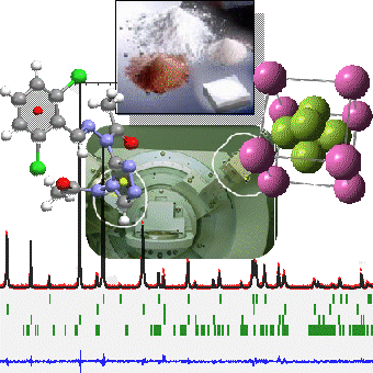
Bragg's work in 1914 determined the first crystal structures (NaCl, KCl, KBr and KI) from diffraction data. The application of X-ray crystallography to the study of the crystalline and molecular structure of structured solids brought the discipline to the attention of the scientific community.
From a technical point of view, in recent years, a large number of accessories have been developed that make a conventional laboratory diffractometer a powerful and highly versatile tool.
Within its multiple applications, the X-ray diffraction technique to study crystalline powder is perhaps the one that has developed the most possibilities of applications not only in the academic field, but also in the industrial field.
Two basic requirements are essential to extract the most information from a conventional crystalline powder diffractogram: the sample must be composed of crystals oriented by chance (avoid preferential orientation), and the crystals must be in sufficiently large numbers to ensure that they present all possible orientations to the incident radiation.
The X-ray Unit has sufficient instruments (agate mortars, internal standards, oriented supports, sieves...) to cover the different analyses offered in reflection geometry. If the user considers it necessary to purchase any other product or material, they will be invoiced for its value or the value of the material consumed.
On the other hand, and due to the latest advances in optical devices and accessories, it is possible to analyse non-powdery systems of samples that do not allow the Bragg-Brentano geometry to be “respected”, so that we can analyse irregular samples, liquids, polymeric fibres, layers...
Users have access to the results interpretation/processing programmes installed on the unit's computers. In addition, the unit has crystallographic databases available for those who need to perform any phase identification.
Commercial software licensed to the X-Ray Unit can be used (e.g. EVA, HIGHScorePlusPlus): EVA, HIGHScorePlus, SAXSAnalysis) to handle the different crystallographic databases available (CSD, PDF-2, ICSD, COD...).
For advanced users, in ab-initio structural resolution, Rietveld refinement, layer analysis, high resolution, SAXS, GISAXS...) software licences are available.
Training courses are offered for the handling, processing and interpretation of data from related experiments.
An important factor in obtaining a quality diffractogram is particle size. The ideal size of a powder sample should be between 1 and 5 μm, so that we can be sure that at positions slightly different from the 2 Θ of the peak maximum point, there will be destructive interference, and lattice tensions will be minimised, thus generating high and narrow peaks. The ideal sample will have to be homogeneous, with the crystals randomly distributed.
As far as the minimum sample quantity is concerned, it will depend on its diffraction power. For samples with high electron density and well crystallised elements, 0.1 g may be sufficient. In general, we should use as little of the sample as possible, trying to avoid pseudo-extinctions due to packing (absorption problems).
Having multi-layer optics on the Siemens, for samples that cannot be pulverised, these will be placed directly at the zero-angle position of the goniometer, and therefore, the area of the sample to be analysed must be specified in detail.
Crystalline powder diffraction can be applied as a complement to the resolution of problems in different types of analysis.
Analysis of ceramic materials
Ceramic material is mostly made up of microcrystalline phases. Using this technique, we can make qualitative and quantitative determinations of the component phases. The manufacturing technology and the origin of the raw materials used can be established through this characterisation. Analyses can be carried out either with a ground sample or without destroying it, as there is the option of working with parallel beam optics. In addition, the temperature chamber will allow us to simulate, in the laboratory, the working conditions in the oven where the part to be analysed was manufactured, controlling the temperature and the working atmosphere (oxidising, reducing).
Analysis of metals and alloys
This technique is sensitive to the compositions of metal alloys. In addition, lattice imperfections caused by trace elements incorporated in the crystallographic lattices of the metal matrix can be determined. The sample can be analysed without destruction.
Analysis of stones and minerals
The chemical and mineralogical composition of a typical mineral can be determined. The analysis may be qualitative, semi-quantitative or quantitative, depending on the desired information.
Analysis of paints
Pigments contained in paints can be analysed by X-ray diffraction. Irregular surfaces can be analysed without prejudice to the data obtained. Depending on the incident angle of the X-ray radiation, only the surface layers can be accessed.
Soil and sediment analysis
Analytical determination (mineralogical analysis, amorphous fraction...).
The mineralogy of the soil is known using this technique, which is essential to determine the origin, age, evolution, physical behaviour, nutrient and contaminant retention capacity, potential fertility...
Especially important is the study of soil clays, which are responsible for its main properties. To facilitate identification, it may be necessary to make an oriented aggregate. This is achieved by dispersing a soil sample or a specific particle size fraction (e.g. less than 50 μ) in water and ammonia. Subsequently, it is expanded on a flat glass where the solvent will evaporate.
The recrystallisation will favour a preferential orientation (majority growth in a certain crystallographic plane), increasing the diffraction power of certain key peaks for its identification. Other treatments such as saturation in different cations and in ethylene glycol or calcination at 550 °C of the oriented aggregates are also mentioned.
Another available application is to know the degree of crystallinity of the mineral components, as well as the presence of amorphous or quantitative compositions of the constituent clays.
This is probably the best-known application of crystal powder diffraction, since the diffractogram obtained is characteristic of the crystalline solid under study. The characteristic values of the peak positions and relative intensities depend on the dimensions of the cell, the microcrystals unit, as well as the nature and distribution of the atoms in three-dimensional and periodic space.
In the 1930s, a relatively simple procedure was devised to identify crystalline solids by comparing diffractograms of measured standards (Hanawalth and Rinn method). Today, the ICDD-JCPDS has collected thousands (>64000) of diffractograms classified according to their nature, easily accessible by specific software. We have access to different updated databases that allow us to develop the required searches.
The analysis of the shape of the peaks in the diffractogram started in the 1980s and allows the imperfections of crystalline solids to be studied. The most relevant information, in general, tends to be the size of the microcrystal and the reticular distortions.
Nowadays, there are many models that allow to the distribution of crystalline domains to be determined by studying the peak profile. Differences between crystalline domain size and reticular distortions can be revealed by different methods of analysis. It is advisable to search literature and see which method is used for the system to be analysed, as different models can interpret the same system, but the models do not tend to give very comparable values. We use single peak methods (e.g.: Scherrer) and global analysis (e.g.: Warren and Averbach), depending on how precise or comparative you want to be.
Since the publication of Hugo Rietveld's work in 1969, there has been a tendency to analyse the diffractogram as type yi(2Θ) functions, an experimental function which allows the intensities of the calculated reflections Ihkl to be distributed in the appropriate diffractogram range. This approach made it possible to deal with peak overlapping in a much more efficient and automatic way (computers), revolutionising the field of diffractogram interpretation. Originally, this method was designed to refine crystal structures. Today, it has been extended to deal with a large number of problems with X-ray data obtained in the laboratory such as:
- Refinement of crystal structures (including complex phase mixtures).
- Quantitative analysis of crystalline phases and the amount of the amorphous phase.
- Resolution of crystal structures ab initio (without prior structural knowledge).
- Estimation of the size and shape of microparticles (coherent diffraction domains).
- Microstructural analysis.
It must be taken into account that in order to obtain a good fit, it is necessary to work with data that has a minimum quality. This is achieved by optimising data collection and if the microcrystals to be analysed are of a suitable size. Therefore, each time these analyses are proposed, it will be decided whether they are feasible or not, depending on the crystallinity or quality of the sample.
The values of the unit cell parameters of the crystalline phases included in the sample to be analysed are obtained from the position of the diffractogram peaks. The hkl indices, i.e. the values of the interplanar spacings of the observed structure, are assigned to each of the observed peaks. There will therefore be a complex relationship between the dhkl and the parameters describing the crystalline network: a, b, c, α, β and γ. We will use different programs to automatically assign the indices. Although all of them require a minimum quality of the data used.
The unit cell will be more reliable the higher the number of observed reflections interpreted. There are different parameters that allow an assessment of the proposed cells (M20, FN), measuring both the accuracy of the observed angular positions and the probability that the proposed cells are correct.
There are different types of quantitative analysis based on X-ray diffraction. The most widely used, historically, use the relative values of integrated intensities of certain characteristic peaks of the component phases. A semi-quantitative estimate of the phases present can be made with the correction applied to each phase (correcting for absorption, diffraction power...), and by comparing the relative intensities.
All the peaks of each phase must be used to obtain quantitative values. For this purpose, there are certain programmes that use different estimations or models. The Rietveld methodology is being used as a further alternative in quantitative determination. In addition, in the case of determining the percentage of the amorphous part, a known amount of an internal standard must be included in the problem mixture.
Due to the difficulty or complication of the possible work, there are different rates depending on the analysis required.
X-ray Unit
- Research Support Building (CACTUS)
- Rúa de Constantino Candeira, 1. Campus Vida , 15782Santiago de Compostela
- 881 816 223